https://animalscienceveterinaryresearchcentre.com/synovial-joints-anatomy


Synovial Joints: Structure, Types, Functions and Veterinary Anatomy Notes
Introduction to Synovial Joints
Synovial Joints are specialized joints in the bodies of animals that allow controlled movement between two or more bones. They are among the most important structures studied in Veterinary Anatomy, Gross Anatomy, Osteology, and Arthrology. Walking, running, jumping, chewing, standing, and maintaining body posture depend on the coordinated function of joints, muscles, ligaments, and bones.
Synovial joints are commonly classified as freely movable joints or diarthroses. Their most characteristic feature is the presence of a joint cavity containing synovial fluid. The articulating surfaces of the bones are generally covered by articular cartilage, while a joint capsule surrounds the articulation and helps maintain its stability.
The structure and function of synovial joints differ among cattle, buffaloes, horses, sheep, goats, dogs, and other domestic animals. These differences are associated with locomotion, body weight, limb structure, and species-specific anatomical adaptations.
Understanding Synovial Joints is essential for veterinary students because joint injuries and diseases can cause pain, lameness, reduced mobility, and decreased productivity. This article explains their definition, structure, classification, movements, functions, clinical importance, common disorders, and important veterinary examination questions.
1. Definition of Synovial Joints
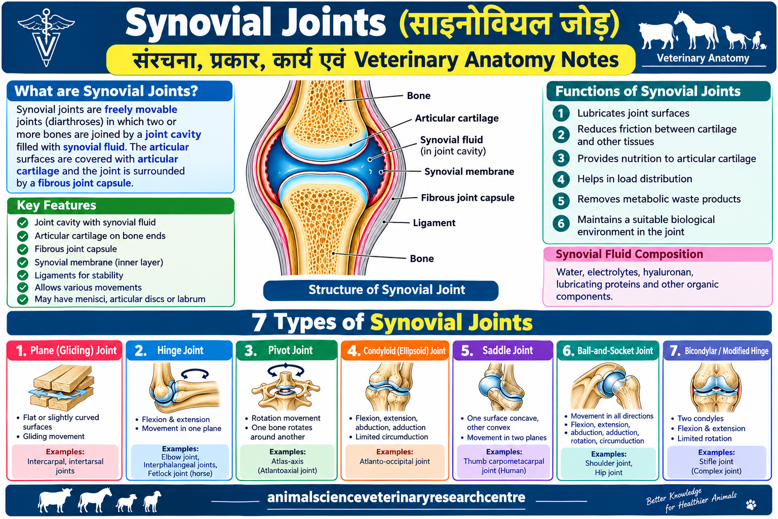
Synovial joints are articulations in which the opposing bone surfaces are separated by a joint cavity containing synovial fluid. The articular surfaces are generally covered by hyaline cartilage, and the joint is enclosed by a capsule consisting of an outer fibrous layer and an inner synovial membrane.
These joints permit different degrees of movement depending on their shape, supporting ligaments, surrounding muscles, and anatomical arrangement.
Examples include the shoulder, elbow, hip, stifle, carpal, and tarsal joints of domestic animals.
Important Characteristics of Synovial Joints
The major characteristics of Synovial Joints are:
- A joint cavity is present between the articulating surfaces.
- Synovial fluid is present within the cavity.
- Articular cartilage covers most opposing bony surfaces.
- A fibrous joint capsule surrounds the articulation.
- A synovial membrane lines the inner surface of the capsule where cartilage is absent.
- Ligaments provide support and help control movement.
- Different joints permit different ranges and directions of movement.
- Some joints contain additional structures such as menisci, articular discs, or labra.
The combination of these structures allows joints to balance mobility, stability, and load-bearing capacity. Synovial Joints: Structure, Types, Functions and Veterinary Anatomy Notes
2. Structure of Synovial Joints
The structure of Synovial Joints is adapted to reduce friction, distribute forces, and permit smooth movement. Each component contributes to the normal functioning of the joint.
2.1 Articular Cartilage
Articular cartilage is a specialized, smooth layer of cartilage covering the articulating surfaces of bones in most synovial joints. It is generally composed of hyaline cartilage.
Its extracellular matrix contains water, collagen, proteoglycans, and other molecules that provide resilience and resistance to compression.
Functions of articular cartilage:
- Reduces friction between opposing joint surfaces.
- Distributes mechanical loads across the joint.
- Protects the underlying bone from excessive stress.
- Provides a smooth surface for movement.
- Helps absorb and transmit forces during locomotion.
Articular cartilage does not contain blood vessels or nerves under normal conditions. Nutrients reach it primarily through diffusion from synovial fluid, with contributions from the underlying tissues to deeper regions.
Damage to articular cartilage can interfere with normal joint movement and may contribute to osteoarthritis. Because cartilage has limited intrinsic repair capacity, significant injuries may have long-term consequences. Synovial Joints: Structure, Types, Functions and Veterinary Anatomy Notes
2.2 Joint Cavity
The joint cavity is the space between the articulating surfaces of a synovial joint. It contains synovial fluid and allows the opposing surfaces to move relative to each other.
The shape and extent of the cavity vary according to the anatomical arrangement of the joint. Some joints have relatively simple cavities, whereas others contain additional structures that modify the internal arrangement.
The joint cavity is an essential distinguishing feature of synovial joints. It provides the environment in which lubrication and the exchange of substances between synovial fluid and articular cartilage occur.
2.3 Joint Capsule
The joint capsule is a connective tissue envelope surrounding the articulation. It helps maintain the relationship between the bones and contributes to joint stability.
The capsule generally consists of two layers.
Fibrous layer: The outer layer is made primarily of dense connective tissue. It provides mechanical strength and helps resist excessive movement.
Synovial membrane: The inner layer covers the internal surfaces of the capsule that are not covered by articular cartilage. It participates in regulating synovial fluid and maintaining the joint environment.
The thickness and strength of the joint capsule vary between joints. Capsules associated with weight-bearing or highly mobile joints are adapted to their particular mechanical requirements.
2.4 Synovial Membrane
The synovial membrane is a specialized connective tissue membrane lining the inner surface of the joint capsule, except over the articular cartilage.
It contains specialized cells called synoviocytes. These cells contribute to the maintenance of synovial fluid and the removal of cellular debris.
Two functional categories of synoviocytes are commonly described:
Type A synoviocytes: These cells have macrophage-like functions and help remove debris and other particles from the joint environment.
Type B synoviocytes: These cells contribute to the production of hyaluronan and other lubricating components of synovial fluid.
The synovial membrane is important in maintaining joint homeostasis. Inflammation of this membrane, known as synovitis, can alter synovial fluid composition and contribute to pain, swelling, and restricted movement.
2.5 Synovial Fluid Synovial Joints: Structure, Types, Functions and Veterinary Anatomy Notes
Synovial fluid is a viscous biological fluid present within the joint cavity. It is usually clear to pale yellow in a healthy joint, although normal characteristics vary with species and joint conditions.
Synovial fluid is related to plasma filtration and is modified by the activity of synovial tissues. Its components include water, electrolytes, hyaluronan, lubricating proteins, and other biological substances.
Functions of synovial fluid:
- Lubricates the articulating surfaces.
- Reduces friction during movement.
- Helps supply nutrients to articular cartilage.
- Supports the removal and transport of metabolic waste products.
- Contributes to load distribution and the mechanical behavior of the joint.
- Helps maintain a suitable biological environment within the joint.
Hyaluronan contributes significantly to the viscosity and viscoelastic properties of synovial fluid. Lubricin and other molecules also help reduce friction at the joint surfaces.
The amount and composition of synovial fluid vary according to the joint, animal species, and physiological condition.
An abnormal accumulation of fluid is called joint effusion. It may occur following injury, inflammation, infection, or other joint disorders.
2.6 Ligaments
Ligaments are strong bands of connective tissue that usually connect one bone to another. They help stabilize joints and restrict excessive or abnormal movement.
Ligaments may be classified according to their anatomical relationship with the joint capsule.
- Capsular ligaments: Thickened regions of the joint capsule.
- Extracapsular ligaments: Ligaments located outside the capsule.
- Intracapsular ligaments: Ligaments situated within the capsule, some of which are separated from the synovial cavity by synovial membrane.
Ligaments are particularly important in weight-bearing joints of cattle, buffaloes, horses, and other domestic animals.
Injury to a ligament may cause pain, instability, abnormal joint movement, and lameness. The severity depends on the affected ligament and the extent of the damage.
2.7 Menisci Synovial Joints: Structure, Types, Functions and Veterinary Anatomy Notes
Menisci are fibrocartilaginous structures present in certain synovial joints. They improve the relationship between articulating surfaces and help distribute mechanical loads.
The stifle joint of domestic animals contains medial and lateral menisci associated with the femorotibial articulation.
Menisci contribute to joint stability, load transmission, and the distribution of pressure across the articular surfaces. Damage to a meniscus may interfere with normal joint mechanics and contribute to lameness.
2.8 Articular Discs
Articular discs are fibrocartilaginous structures located between articulating surfaces in certain joints. Depending on the joint, a disc may divide the joint cavity into separate compartments.
They help improve the fit between surfaces and distribute forces during movement.
Articular discs should not be confused with menisci, although both are fibrocartilaginous structures that modify joint mechanics.
2.9 Articular Labrum
An articular labrum is a fibrocartilaginous rim attached to the margin of certain joint sockets. It increases the effective depth of the socket and helps improve joint stability.
The glenoid labrum of the shoulder and the acetabular labrum of the hip are well-known examples in human anatomy. Veterinary students should consult species-specific anatomical references when studying equivalent structures in domestic animals.
3. Seven Major Types of Synovial Joints
Synovial joints are classified according to the shape of their articular surfaces and the movements they permit. A commonly taught classification includes plane, hinge, pivot, condyloid, saddle, ball-and-socket, and bicondylar or modified hinge joints.
The exact classification of some complex joints may vary between anatomical textbooks.
3.1 Plane or Gliding Joint
Plane joints have relatively flat or slightly curved articular surfaces. They primarily permit limited sliding or gliding movements.
Examples:
- Intercarpal joints.
- Many intertarsal articulations.
- Certain small articular joints between vertebral processes.
Plane joints generally have a limited range of movement individually. However, the combined movement of several joints may contribute significantly to the flexibility of a region.
Main movements: Gliding and limited translation.
Veterinary importance: Intercarpal and intertarsal joints contribute to the coordinated movement of the limbs. Their function is important during standing and locomotion.
Key examination fact: Plane joints are also called gliding joints.
3.2 Hinge Joint
Hinge joints primarily permit flexion and extension. Their movement resembles the opening and closing of a hinge, although biological joints are more complex than mechanical hinges.
Examples:
- Elbow joint, which behaves predominantly as a hinge-like articulation.
- Interphalangeal joints.
- The equine fetlock joint, where flexion and extension are major movements.
Hinge-like joints allow the limbs to bend and straighten during walking, running, and standing. Their stability depends on the articular surfaces, capsule, ligaments, and surrounding muscles.
Main movements: Flexion and extension.
Veterinary importance: Hinge-like joints are essential for coordinated limb movement. Injury or inflammation may restrict motion and produce lameness.
Key examination fact: Flexion and extension are the principal movements of a hinge joint.
3.3 Pivot Joint
Pivot joints permit rotation around an axis. One bone rotates relative to another bone or a supporting ligamentous arrangement.
Example:
The atlantoaxial articulation between the atlas and axis allows important rotational movements of the head and neck.
In some species, the proximal radioulnar articulation is described as pivot-like. However, the arrangement and range of rotation of the radius and ulna differ among domestic animals.
Main movement: Rotation.
Veterinary importance: Pivot-like articulations help provide rotational movement where the anatomical arrangement permits it.
Key examination fact: Rotation is the principal movement of a pivot joint.
3.4 Condyloid or Ellipsoid Joint
Condyloid joints have an oval or ellipsoid articular surface that articulates with a complementary surface.
They may permit movement in two principal planes, including flexion, extension, abduction, and adduction. Circumduction may occur as a combination of these movements.
The atlanto-occipital articulation is commonly used as an example of an ellipsoid-like joint in comparative anatomy. It contributes to flexion and extension of the head, along with some lateral bending.
Main movements: Flexion, extension, abduction, adduction, and limited circumduction, depending on the joint.
Veterinary importance: Understanding the shape of articular surfaces helps explain how joints move and how their ligaments limit excessive movement.
Key examination fact: Condyloid joints have oval or ellipsoid articular surfaces.
3.5 Saddle Joint
In a saddle joint, each articulating surface is concave in one direction and convex in another. The two surfaces fit together in a saddle-like arrangement.
This structure allows controlled movement in more than one plane.
A classic example is the carpometacarpal joint of the human thumb. This example is useful for comparative anatomy, but it should not automatically be applied to domestic animals without species-specific evidence.
Main movements: Commonly biaxial movements, with the exact range depending on the articulation.
Veterinary importance: Saddle-shaped articular surfaces illustrate how joint geometry influences movement and stability.
Key examination fact: A saddle joint is concave in one direction and convex in another.
3.6 Ball-and-Socket Joint
Ball-and-socket joints contain a rounded articular head that fits into a cup-like socket. These joints allow movement in several directions.
Examples:
- Shoulder joint.
- Hip joint.
The shoulder joint generally allows a relatively wide range of movement. The hip joint is usually more constrained by its socket, ligaments, and surrounding tissues, which contribute to stability.
Main movements:
- Flexion.
- Extension.
- Abduction.
- Adduction.
- Rotation.
- Circumduction.
The extent of each movement varies among species and depends on the shape of the articular surfaces and supporting structures.
Veterinary importance: These joints are essential for limb positioning and movement. Their disorders may cause pain, instability, and lameness.
Key examination fact: The shoulder and hip are major examples of ball-and-socket joints.
3.7 Bicondylar or Modified Hinge Joint
Bicondylar joints contain two prominent condylar articular regions. Many descriptions of complex joints emphasize their predominantly flexion-extension movement, together with limited rotation or other accessory movements.
The stifle joint of domestic animals is a complex synovial articulation that includes femorotibial and femoropatellar components.
The menisci, patella, joint capsule, and multiple ligaments contribute to its function.
Main movements: Primarily flexion and extension, with limited accessory movements depending on the anatomical structure.
Veterinary importance: The stifle is clinically important in horses, cattle, dogs, and other animals. Injury to its ligaments or menisci can impair locomotion.
Key examination fact: The stifle is a complex synovial joint, often described as modified hinge-like.
Some anatomical classifications list bicondylar joints separately, while others classify particular examples as modified hinge or complex synovial joints. Follow the classification used in your veterinary anatomy syllabus.
4. Movements of Synovial Joints
The movements of Synovial Joints depend on the shape of the articulating surfaces, the arrangement of ligaments, and the action of muscles.
4.1 Flexion
Flexion generally decreases the angle between the bones forming a joint. For example, bending the elbow is a flexion movement.
In domestic animals, flexion is important for limb advancement, lying down, rising, and adjusting body posture.
4.2 Extension
Extension generally increases the angle between the bones or straightens a flexed joint.
It is important during limb support, standing, and the propulsive phases of locomotion.
4.3 Abduction
Abduction is the movement of a body part away from the median plane.
The permitted range depends on the particular joint and the restrictions imposed by its ligaments and surrounding structures.
4.4 Adduction
Adduction is the movement of a body part toward the median plane.
Abduction and adduction are most evident in joints that permit movement away from and toward the body’s midline.
4.5 Rotation
Rotation occurs when a bone turns around its longitudinal axis.
The atlantoaxial articulation contributes to rotational movement of the head. The amount of rotation possible at other joints depends on their structure.
4.6 Circumduction
Circumduction is a combined movement in which the distal part of a limb describes a circular or cone-shaped path.
It combines flexion, extension, abduction, and adduction. Its range varies among joints and animal species.
4.7 Gliding
Gliding occurs when one articular surface slides over another with limited translation.
This movement is particularly important in plane joints and in the combined function of small carpal and tarsal articulations.
5. Functions of Synovial Joints
Synovial joints perform several essential functions in the animal body.
5.1 Facilitation of Movement
They allow bones to move relative to one another, making walking, running, jumping, feeding, and other activities possible.
5.2 Weight Bearing
Many synovial joints help transmit forces between the limbs and the trunk. Their structures distribute loads and contribute to postural support.
5.3 Reduction of Friction
Articular cartilage and synovial fluid help reduce friction between the opposing joint surfaces.
5.4 Shock and Load Management
The cartilage, fluid, and supporting tissues work together to distribute forces and moderate local mechanical stress.
5.5 Maintenance of Stability
Ligaments, joint capsules, articular geometry, and surrounding muscles help prevent excessive or abnormal movements.
5.6 Support of Locomotion
Healthy joints are essential for normal gait. Joint pain, swelling, or instability can alter stride length, limb loading, and overall mobility.
5.7 Contribution to Daily Activities
In livestock, joint function affects grazing, feeding, walking, standing, reproduction-related activity, and access to water. In working and athletic animals, it is also important for performance.
6. Synovial Fluid: Composition and Clinical Importance
Synovial fluid is essential for the maintenance of healthy synovial joints. Its functions extend beyond lubrication.
Composition
Synovial fluid contains:
- Water.
- Electrolytes.
- Hyaluronan.
- Lubricating proteins such as lubricin.
- Small quantities of nutrients and other biological molecules.
- Cellular components that may vary with the condition of the joint.
Hyaluronan contributes to viscosity, while lubricin helps reduce friction at articulating surfaces.
Lubrication
Synovial fluid reduces friction between cartilage surfaces during movement. Its lubricating properties help protect the articular surfaces from excessive wear.
Nutrition of Articular Cartilage
Because articular cartilage lacks a direct blood supply under normal conditions, nutrients reach chondrocytes primarily through diffusion from synovial fluid and related tissue exchanges.
Waste Transport
Synovial fluid supports the transport of metabolic waste products away from cartilage and other joint tissues.
Joint Homeostasis
Normal synovial fluid helps maintain a suitable environment for the cells and tissues within the joint.
Abnormal Synovial Fluid
Injury, inflammation, and infection can alter synovial fluid volume, appearance, viscosity, and cellular composition.
In septic arthritis, inflammatory changes may be marked. However, fluid appearance alone cannot reliably confirm or exclude infection. Veterinary examination and appropriate laboratory investigations are needed.
7. Synovial Joints in Domestic Animals
The anatomy and clinical importance of Synovial Joints differ according to species, locomotor requirements, and limb structure.
7.1 Cattle and Buffaloes
Cattle and buffaloes rely on healthy joints for standing, walking, grazing, feeding, and carrying body weight.
Important joints include:
- Shoulder joint.
- Elbow joint.
- Carpal joints.
- Stifle joint.
- Tarsal joints.
- Fetlock and interphalangeal joints.
Lameness can reduce feed intake, mobility, productivity, and reproductive performance. Hoof health, suitable flooring, nutrition, and early recognition of joint disorders are important aspects of herd management.
7.2 Horses
Horses require healthy joints for walking, trotting, cantering, and running.
Important joints include the shoulder, elbow, carpus, fetlock, interphalangeal joints, and stifle.
The distal limb joints of horses experience substantial mechanical loading during locomotion. Injury or inflammation in these regions may cause lameness and reduced athletic performance.
Regular assessment, appropriate exercise, suitable footing, and prompt veterinary care are important for joint health.
7.3 Dogs
In dogs, synovial joints enable running, jumping, playing, and everyday movement.
Important joints include the shoulder, elbow, hip, stifle, carpus, and tarsus.
Common clinical problems include osteoarthritis, hip dysplasia, elbow dysplasia, and cranial cruciate ligament injury. These disorders may produce pain, stiffness, instability, or altered gait.
Body-weight management, appropriate activity, and veterinary assessment can help support joint health.
7.4 Sheep and Goats
Sheep and goats depend on healthy joints for walking, grazing, climbing, and supporting body weight.
Carpal, tarsal, stifle, and digit joints are important in locomotion. Hoof care, balanced nutrition, suitable housing, and early treatment of lameness help maintain mobility.
7.5 Comparative Veterinary Importance
Although the fundamental principles of synovial joint anatomy are shared across domestic mammals, specific bone shapes, ligament arrangements, and permitted movements may differ considerably.
Veterinary students should therefore combine general anatomical principles with species-specific descriptions when preparing for examinations or clinical practice.
8. Common Diseases and Disorders of Synovial Joints
Synovial joints can be affected by infection, inflammation, degeneration, trauma, and developmental abnormalities.
8.1 Arthritis
Arthritis is a general term for inflammation of a joint. It may occur because of infection, immune-mediated disease, trauma, or other conditions.
Possible signs include:
- Joint swelling.
- Pain or sensitivity.
- Increased local warmth.
- Lameness.
- Reduced range of motion.
- Difficulty bearing weight.
Diagnosis depends on clinical examination and, where appropriate, imaging and laboratory tests.
8.2 Septic Arthritis
Septic arthritis is an infection within a joint. Microorganisms may enter through penetrating injuries, contaminated wounds, surgical procedures, or the bloodstream.
Young animals may develop joint infections associated with systemic infections.
Septic arthritis is potentially destructive because inflammation and infection can rapidly damage articular cartilage.
Prompt veterinary assessment is essential. Depending on the case, treatment may include appropriate antimicrobial therapy, joint lavage, and supportive care.
8.3 Osteoarthritis
Osteoarthritis is a degenerative joint disorder involving changes in articular cartilage, subchondral bone, and other joint tissues.
Possible signs include chronic lameness, stiffness after rest, reduced mobility, and discomfort following exercise.
Risk factors and progression vary among species and individuals. Management may include veterinary-directed pain control, weight management, appropriate exercise, rehabilitation, and other treatments selected for the animal.
8.4 Joint Effusion
Joint effusion refers to an abnormal accumulation of fluid within a joint.
It may be associated with inflammation, injury, infection, or other joint diseases. Visible swelling alone does not identify the underlying cause.
Veterinary examination is required to determine the cause and appropriate management.
8.5 Ligament Injuries
Ligament injuries may range from mild stretching to partial or complete tearing.
In dogs, cranial cruciate ligament injury is an important cause of stifle instability and lameness.
The clinical presentation depends on the ligament involved and the severity of the injury. Diagnosis may require orthopedic examination and imaging.
8.6 Developmental Joint Disorders
Developmental joint disorders arise when normal development of a joint or its associated structures is disrupted.
Hip dysplasia and elbow dysplasia are important examples in dogs. Genetic factors, growth, body condition, and other influences may affect disease risk.
Early veterinary assessment can help determine suitable monitoring and management.
9. Clinical Examination of Synovial Joints
A systematic joint examination is an important part of veterinary clinical practice.
9.1 Observation
The animal’s posture, gait, weight-bearing ability, and visible swelling are assessed.
Gait observation can help identify which limb is affected and how movement has changed.
9.2 Palpation
The veterinarian carefully examines the joint for pain, heat, swelling, fluid accumulation, and abnormal structures.
Palpation should be performed with care, particularly when a fracture or severe injury is suspected.
9.3 Range-of-Motion Assessment
The joint is assessed for its available movement, stiffness, discomfort, and abnormal restriction.
Forceful movement should be avoided when severe injury or instability is suspected.
9.4 Radiography
Radiographs help evaluate bone structure, fractures, joint alignment, and some degenerative changes.
Radiography is useful in many orthopedic investigations, although it does not directly show all soft-tissue abnormalities.
9.5 Ultrasound and Advanced Imaging
Ultrasound can help assess selected soft tissues, fluid accumulation, and accessible joint structures.
CT or MRI may be appropriate in selected cases when more detailed evaluation is required.
9.6 Synovial Fluid Analysis
When indicated, a veterinarian may collect synovial fluid for analysis.
Testing may include assessment of appearance, cellular composition, and other laboratory characteristics. Bacterial culture may be indicated when infection is suspected.
Joint aspiration should be performed by trained veterinary personnel using appropriate aseptic technique.
Clinical warning: A painful, swollen joint, sudden severe lameness, or joint swelling accompanied by fever requires prompt veterinary attention. Do not administer medication or inject a joint without veterinary guidance.
10. Differences Between Synovial, Fibrous, and Cartilaginous Joints
Joints are commonly classified structurally as fibrous, cartilaginous, or synovial.
Fibrous Joints
In fibrous joints, bones are connected by fibrous connective tissue. Movement is generally absent or very limited.
Skull sutures are a classic example.
Cartilaginous Joints
In cartilaginous joints, bones are connected by cartilage. The amount of movement depends on the type and structure of the articulation.
Examples include intervertebral symphyses and certain cartilaginous articulations.
Synovial Joints
Synovial joints contain a joint cavity and synovial fluid. They generally permit more movement than fibrous or cartilaginous joints.
Examples include the shoulder, elbow, hip, stifle, and many carpal and tarsal joints.
The presence of a joint cavity is the key structural feature distinguishing synovial joints from the other major joint categories.
11. Prevention and Maintenance of Joint Health in Animals
Maintaining joint health is important for animal welfare, mobility, and productivity.
Recommended general measures include:
- Provide a balanced diet appropriate for the species, age, and physiological condition.
- Maintain a suitable body condition and avoid excessive weight gain.
- Provide appropriate exercise and avoid sudden increases in physical workload.
- Reduce the risk of slips, falls, and traumatic injuries.
- Maintain suitable flooring and housing conditions.
- Inspect hooves and limbs regularly in livestock.
- Recognize lameness, swelling, or reduced mobility early.
- Seek veterinary assessment when joint disease is suspected.
- Follow appropriate hygiene and infection-control practices.
- Follow veterinary recommendations for rehabilitation and recovery after injury.
These measures can help reduce certain risk factors but cannot prevent every joint disease.
12. Important Veterinary Anatomy Examination Facts
- Synovial joints are commonly described as freely movable joints or diarthroses.
- The joint cavity is a defining feature of synovial joints.
- Articular cartilage is generally hyaline cartilage.
- The synovial membrane lines the inner joint capsule where articular cartilage is absent.
- Hyaluronan contributes to the viscosity of synovial fluid.
- Lubricin contributes to joint lubrication.
- Ligaments connect bones and help stabilize joints.
- Plane joints primarily permit gliding movements.
- Hinge joints primarily permit flexion and extension.
- Pivot joints primarily permit rotation.
- Ball-and-socket joints allow movement in several directions.
- The shoulder and hip are major ball-and-socket joint examples.
- Menisci are fibrocartilaginous structures.
- Joint effusion means an abnormal accumulation of fluid within a joint.
- Septic arthritis is a potentially serious joint infection.
- Osteoarthritis involves degenerative changes in joint tissues.
- Synovial fluid analysis can help investigate selected joint disorders.
- The exact classification of some complex joints varies between anatomical references.
13. Important MCQs on Synovial Joints
Question 1. What is the defining structural feature of a synovial joint?
A. Complete absence of a joint cavity
B. Presence of a joint cavity
C. Connection exclusively by fibrous tissue
D. Absence of articular surfaces
Correct Answer: B. Presence of a joint cavity
Explanation: Synovial joints contain a joint cavity between the articulating surfaces, and this cavity contains synovial fluid.
Question 2. Which substance contributes significantly to the viscosity of synovial fluid?
A. Hemoglobin
B. Hyaluronan
C. Keratin
D. Myosin
Correct Answer: B. Hyaluronan
Explanation: Hyaluronan contributes to the viscoelastic properties of synovial fluid and helps support lubrication.
Question 3. Which is a major example of a ball-and-socket joint?
A. Shoulder joint
B. Skull suture
C. Tooth socket
D. Fibrous joint
Correct Answer: A. Shoulder joint
Explanation: The shoulder is a ball-and-socket joint that permits movement in multiple directions.
Question 4. Which movements are characteristic of hinge-like joints?
A. Rotation only
B. Flexion and extension
C. Gliding only
D. Circumduction only
Correct Answer: B. Flexion and extension
Explanation: Hinge-like joints primarily allow bending and straightening movements.
Question 5. Which structure connects bones and helps stabilize joints?
A. Ligament
B. Hair follicle
C. Epidermis
D. Tooth enamel
Correct Answer: A. Ligament
Explanation: Ligaments connect bones and help control excessive or abnormal joint movements.
Question 6. What is a major function of articular cartilage?
A. Producing blood cells
B. Reducing friction and distributing loads
C. Producing bile
D. Secreting digestive enzymes
Correct Answer: B. Reducing friction and distributing loads
Explanation: Articular cartilage provides a smooth surface and helps distribute forces during joint movement.
Question 7. What is septic arthritis?
A. Infection within a joint
B. A disorder limited to hair follicles
C. A condition affecting only the skin
D. A digestive disorder
Correct Answer: A. Infection within a joint
Explanation: Septic arthritis involves infection and inflammation within a joint and requires prompt veterinary evaluation.
Question 8. What does joint effusion mean?
A. Complete fusion of two bones
B. Abnormal accumulation of fluid in a joint
C. Complete loss of a ligament
D. Formation of a new tendon
Correct Answer: B. Abnormal accumulation of fluid in a joint
Explanation: Joint effusion may occur with inflammation, trauma, infection, or other joint disorders.
Question 9. Which type of joint primarily permits gliding movement?
A. Plane joint
B. Pivot joint
C. Ball-and-socket joint
D. Saddle joint
Correct Answer: A. Plane joint
Explanation: Plane joints have relatively flat articular surfaces that permit limited sliding movements.
Question 10. Which condition is characterized by degenerative changes in joint tissues?
A. Osteoarthritis
B. Rhinitis
C. Gastritis
D. Dermatitis
Correct Answer: A. Osteoarthritis
Explanation: Osteoarthritis affects articular cartilage and other joint tissues and may cause chronic pain and restricted movement.
14. Frequently Asked Questions About Synovial Joints
Q1. What are Synovial Joints?
Synovial joints are articulations containing a joint cavity filled with synovial fluid. They generally permit controlled movement between the articulating bones.
Q2. What is the main function of synovial fluid?
Synovial fluid lubricates joint surfaces, supports the nutrition of articular cartilage, and helps maintain a suitable environment within the joint.
Q3. How many types of synovial joints are commonly described?
A commonly taught classification includes seven categories: plane, hinge, pivot, condyloid, saddle, ball-and-socket, and bicondylar or modified hinge. Classification may vary between textbooks.
Q4. What type of cartilage covers most synovial joint surfaces?
Most articular surfaces in synovial joints are covered by hyaline cartilage.
Q5. Why can synovial joints become painful and swollen?
Possible causes include trauma, arthritis, infection, osteoarthritis, ligament injury, and other joint disorders. Veterinary examination is needed to identify the cause.
Q6. Can all synovial joint diseases be treated in the same way?
No. Treatment depends on the underlying cause, affected species, age, severity, and the specific joint involved.
Q7. Why are synovial joints important in veterinary medicine?
They are essential for locomotion, posture, weight bearing, and normal daily activity. Joint disorders can cause lameness, pain, reduced mobility, and loss of productivity.
Conclusion
Synovial Joints are essential components of the musculoskeletal system of domestic animals. Their joint cavity, synovial fluid, articular cartilage, synovial membrane, capsule, and ligaments work together to support movement, lubrication, stability, and load distribution.
Understanding the seven commonly taught types of synovial joints helps veterinary students connect joint structure with function. Knowledge of synovial fluid, joint movements, species-specific anatomy, and common joint disorders is valuable for veterinary examinations and clinical practice.
Early recognition of joint pain, swelling, instability, and lameness is important for animal welfare. Appropriate veterinary diagnosis and treatment help guide the management of joint disease.
References and Further Reading
- Merck Veterinary Manual: https://www.merckvetmanual.com/
- OpenStax Anatomy and Physiology: https://openstax.org/details/books/anatomy-and-physiology-2e
- Animal Science & Veterinary Research Centre: https://animalscienceveterinaryresearchcentre.com/
Image SEO Details
Recommended image file name: synovial-joints-veterinary-anatomy.jpg
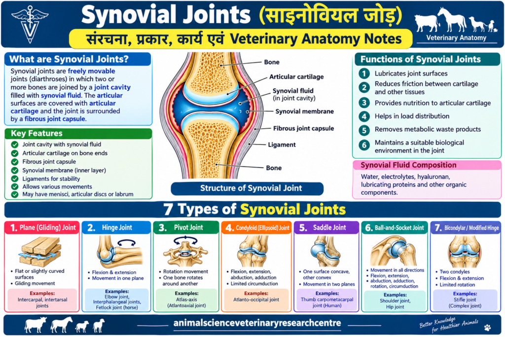
Image Alt Text: Synovial Joints structure showing synovial fluid, articular cartilage, synovial membrane, and joint capsule.
Image Caption: Structure and seven major types of Synovial Joints in Veterinary Anatomy.
Internal Linking Suggestions
Add links to relevant, published articles on your website, such as:
- Veterinary Anatomy: Complete Notes and Diagrams
- Gross Anatomy: Veterinary Study Notes
- Osteology: Animal Skeletal System
- Arthrology: Classification and Structure of Joints
- Histology: Cartilage and Connective Tissue
Use the actual URLs of your published pages. Do not create links to pages that do not exist.
Final SEO Checklist
- Set the Focus Keyword to Synovial Joints in Rank Math.
- Include the keyword near the beginning of the SEO title.
- Add the meta description provided above.
- Set the permalink to
synovial-joints. - Use the Focus Keyword naturally in the opening paragraph and relevant headings.
- Add a labelled anatomical diagram with descriptive alt text.
- Add internal links to related veterinary anatomy articles.
- Retain relevant, trustworthy external references.
- Add a Table of Contents using your WordPress block or plugin.
- Preview the article on mobile and desktop before publishing.
- Check indexing through Google Search Console after publication.
https://teachmeanatomy.info/the-basics/joints-basic/synovial-joint
Synovial Joints: Structure, Types, Functions and Veterinary Anatomy NotesSynovial Joints: Structure, Types, Functions and Veterinary Anatomy NotesSynovial Joints: Structure, Types, Functions and Veterinary Anatomy NotesSynovial Joints: Structure, Types, Functions and Veterinary Anatomy NotesSynovial Joints: Structure, Types, Functions and Veterinary Anatomy NotesSynovial Joints: Structure, Types, Functions and Veterinary Anatomy NotesSynovial Joints: Structure, Types, Functions and Veterinary Anatomy NotesSynovial Joints: Structure, Types, Functions and Veterinary Anatomy NotesSynovial Joints: Structure, Types, Functions and Veterinary Anatomy NotesSynovial Joints: Structure, Types, Functions and Veterinary Anatomy NotesSynovial Joints: Structure, Types, Functions and Veterinary Anatomy NotesSynovial Joints: Structure, Types, Functions and Veterinary Anatomy Notes
1 thought on “Synovial Joints: Structure, Types, Functions and Veterinary Anatomy Notes”