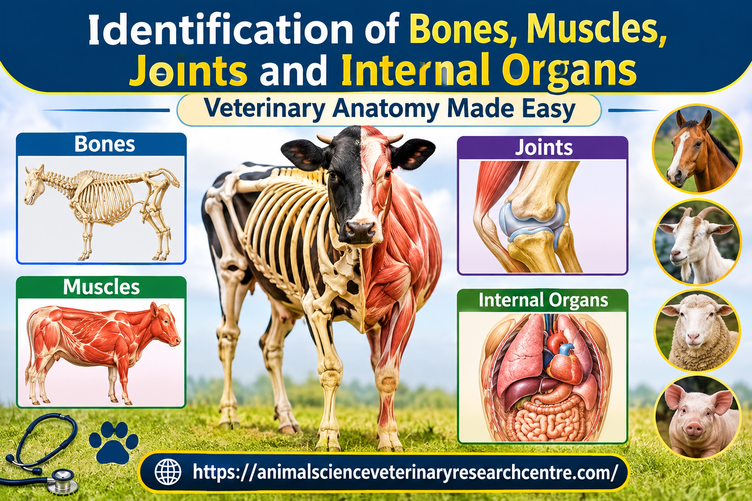


Identification of Bones, Muscles, Joints and Internal Organs
Introduction
The Identification of Bones, Muscles, Joints and Internal Organs is an essential part of Veterinary Anatomy. It helps veterinary students, animal science students, livestock professionals and researchers understand the structure, location and functions of different body parts in domestic animals. Correct anatomical identification is important for physical examination, diagnosis, surgery, injection techniques, animal handling and veterinary clinical practice.
Animal anatomy is commonly studied through gross anatomy, histology, comparative anatomy, osteology, myology, arthrology and splanchnology. These branches explain the structure of bones, muscles, joints and internal organs in animals such as cattle, buffalo, sheep, goats, horses, dogs, cats and pigs.
Anatomical identification requires more than memorising names. Students must understand anatomical landmarks, body regions, orientation, tissue characteristics and relationships between organs. Practical learning using skeletons, preserved specimens, anatomical models, radiographs and diagrams makes identification easier and more accurate.
This article explains the basic principles, important structures, identification methods, species differences and clinical applications of veterinary anatomy.
Table of Contents
- Introduction to Veterinary Anatomy
- Anatomical Position and Directional Terms
- Identification of Bones
- Identification of Muscles
- Identification of Joints
- Identification of Internal Organs
- Comparative Anatomy of Domestic Animals
- Practical Methods of Anatomical Identification
- Clinical Applications
- Common Identification Mistakes
- Veterinary Anatomy Practical Checklist
- Frequently Asked Questions
- Conclusion
1. Introduction to Veterinary Anatomy
Veterinary anatomy is the scientific study of the structure and organisation of animal bodies. It describes the location, shape, size, connections and relationships of different structures.
The body can be divided into several major systems, including the skeletal, muscular, digestive, respiratory, circulatory, urinary, reproductive, nervous and endocrine systems.
Major Branches of Veterinary Anatomy
Gross Anatomy: The study of structures visible to the naked eye, including bones, muscles, joints and internal organs.
Osteology: The study of bones, their shapes, landmarks, articulations and functions.
Myology: The study of muscles, including their origins, insertions, actions and nerve supply.
Arthrology: The study of joints and the connections between bones.
Splanchnology: The study of internal organs, particularly those associated with the digestive, respiratory, urinary and reproductive systems.
Histology: The microscopic study of tissues and their cellular organisation.
Comparative Anatomy: The study of anatomical similarities and differences among animal species.
These branches are interconnected. For example, understanding the femur requires knowledge of its bony landmarks, associated muscles, hip and stifle joints, and nearby blood vessels and nerves.
2. Anatomical Position and Directional Terms
Before identifying bones, muscles, joints or organs, students must understand standard anatomical terminology. These terms help describe the exact position of a structure without confusion.
2.1 Important Directional Terms
- Cranial: Toward the head.
- Caudal: Toward the tail.
- Rostral: Toward the nose, especially when describing the head.
- Dorsal: Toward the back or upper surface, depending on the body region.
- Ventral: Toward the belly or lower surface.
- Medial: Toward the median plane of the body.
- Lateral: Away from the median plane.
- Proximal: Closer to the trunk or point of origin of a limb.
- Distal: Farther from the trunk or point of origin of a limb.
- Superficial: Near the body surface.
- Deep: Farther from the body surface.
- Palmar: The caudal surface of the forelimb distal to the carpus.
- Plantar: The caudal surface of the hindlimb distal to the tarsus.
2.2 Anatomical Planes
Median plane: Divides the body into equal right and left halves.
Sagittal plane: Divides the body into right and left portions that may not be equal.
Dorsal plane: Separates dorsal and ventral portions.
Transverse plane: Cuts across the body or a body part, approximately perpendicular to its long axis.
These planes are especially useful when interpreting radiographs, CT scans, MRI images and anatomical sections.
3. Identification of Bones
Bones form the skeletal framework of the body. They support body weight, protect internal organs, provide attachment sites for muscles and participate in movement and blood cell formation.
The adult skeleton of a typical domestic mammal is organised into the axial skeleton and appendicular skeleton, with the exact number of bones varying by species and anatomical counting method.
3.1 Classification of Bones
Bones are classified according to their shape.
Long bones: Longer than they are wide. Examples include the humerus, radius, femur and tibia. They act as levers for movement.
Short bones: Approximately cube-shaped. Examples include many carpal and tarsal bones. They provide stability and distribute forces.
Flat bones: Thin or flattened bones that often protect organs and provide broad muscle attachment areas. Examples include parts of the skull, scapula and ribs.
Irregular bones: Bones with complex shapes, such as vertebrae.
Sesamoid bones: Bones that develop within certain tendons. The patella is a major example.
3.2 Axial Skeleton
The axial skeleton forms the central framework of the body. It includes the skull, vertebral column, ribs and sternum.
Skull: Protects the brain and supports the structures of the face. Important bones include the frontal, parietal, temporal, occipital, maxilla and mandible.
Vertebral column: Consists of cervical, thoracic, lumbar, sacral and caudal vertebrae. The vertebral column protects the spinal cord and supports the trunk.
Ribs: Form the lateral walls of the thorax and protect the heart and lungs.
Sternum: Forms the ventral part of the thoracic skeleton and provides attachment for ribs and muscles.
3.3 Appendicular Skeleton
The appendicular skeleton consists of the bones of the limbs and their supporting structures.
The forelimb includes the scapula, humerus, radius, ulna, carpal bones, metacarpal bones, phalanges and associated sesamoid bones.
The hindlimb includes the pelvic bones, femur, patella, tibia, fibula to varying degrees depending on species, tarsal bones, metatarsal bones, phalanges and sesamoid bones.
3.4 How to Identify a Bone
A systematic approach improves accuracy.
- Determine whether the bone belongs to the axial or appendicular skeleton.
- Classify it by shape.
- Identify its proximal and distal ends.
- Locate distinctive landmarks such as processes, condyles, fossae, foramina and tubercles.
- Identify the articular surfaces.
- Compare the bone with a labelled anatomical diagram.
- Determine its likely species and side, where possible.
For example, the femur is identified by its head, neck, greater and lesser trochanters, shaft and distal condyles. The head articulates with the acetabulum of the pelvis to form the hip joint.
3.5 Important Bony Landmarks
- Process: A projection from a bone.
- Condyle: A rounded articular prominence.
- Fossa: A depression or hollow.
- Foramen: An opening through a bone.
- Tuberosity: A roughened prominence, commonly associated with tendon or ligament attachment.
- Crest: A prominent ridge.
- Trochanter: A large projection found on the femur of many mammals.
Bony landmarks are important for identifying muscle attachments, locating joints and selecting safe sites for certain clinical procedures.
3.6 Clinical Importance of Bone Identification
Bone identification helps in diagnosing fractures, interpreting radiographs, understanding limb deformities and planning orthopaedic surgery. Veterinary students should learn the normal shape and orientation of bones before studying pathological changes.
4. Identification of Muscles
Muscles generate movement, maintain posture, stabilise joints and produce heat. Skeletal muscles are particularly important in locomotion and clinical examination.
4.1 Types of Muscle Tissue
Skeletal muscle: Usually under voluntary control and attached to bones or other structures. It produces body movement.
Smooth muscle: Found in the walls of many hollow organs and blood vessels. It controls movements such as intestinal contractions.
Cardiac muscle: Found in the heart wall. Its specialised structure supports rhythmic contraction.
4.2 Classification of Skeletal Muscles
Muscles may be classified according to their shape, location, action, fibre arrangement or number of heads.
- Fusiform muscles: Spindle-shaped, with a thicker middle and tapered ends.
- Flat muscles: Broad and thin, often forming body walls.
- Circular muscles: Surround openings and act as sphincters.
- Pennate muscles: Have fibres arranged obliquely relative to their tendons.
- Digastric muscles: Have two muscle bellies connected by an intermediate tendon.
4.3 Origin, Insertion and Action
The origin is conventionally the attachment considered more fixed during a particular movement. The insertion is the attachment considered more mobile. These terms depend on the action being described and are not absolute in every movement.
The action is the movement produced by a muscle or muscle group.
Muscles may act as flexors, extensors, abductors, adductors, rotators or sphincters.
For example, the biceps brachii contributes to flexion of the elbow and has additional functions that vary with species and joint position. The quadriceps femoris is the major extensor of the stifle.
4.4 Superficial and Deep Muscles
Superficial muscles lie closer to the skin and can often be recognised during dissection or palpation. Deep muscles lie beneath superficial layers and may act directly on joints or stabilise the vertebral column.
Important muscle groups include:
- Head: Masseter and temporalis, which help close the jaw.
- Neck: Brachiocephalicus and splenius, which contribute to head and neck movements.
- Thoracic limb: Triceps brachii, biceps brachii and the extensor and flexor groups.
- Trunk: Longissimus and abdominal wall muscles.
- Pelvic limb: Gluteal muscles, quadriceps femoris, hamstring group and gastrocnemius.
- Respiratory system: Diaphragm and intercostal muscles.
4.5 How to Identify a Muscle
Begin by locating the muscle’s body region. Identify its fibre direction, attachments, shape and relationship to nearby bones and muscles. Trace its tendon where possible and determine which joint or body part it moves.
A muscle should not be identified by shape alone because neighbouring muscles may look similar. Confirm its attachments and anatomical position using a reliable atlas or dissection guide.
4.6 Clinical Importance of Muscle Identification
Muscle anatomy supports the evaluation of lameness, muscle wasting, trauma and nerve injuries. It also helps veterinary professionals understand intramuscular injection sites, surgical approaches and the effects of muscle damage on movement.
Injection procedures must follow species-specific veterinary guidance because important nerves and blood vessels may lie close to muscle groups.
5. Identification of Joints
A joint, or articulation, is a connection between two or more bones or between bone and cartilage. Joints provide stability and, where appropriate, permit movement.
5.1 Classification of Joints
Joints may be classified structurally or functionally.
Fibrous joints: Bones are connected by dense connective tissue. They permit little or no movement. Many skull sutures are examples.
Cartilaginous joints: Bones are connected by cartilage. They provide support and allow limited movement. Intervertebral connections include cartilaginous components.
Synovial joints: Have a joint cavity containing synovial fluid. They are generally freely movable and are common in the limbs.
5.2 Structure of a Synovial Joint
A typical synovial joint contains:
- Articular cartilage covering the opposing articular surfaces.
- A joint cavity containing synovial fluid.
- A fibrous joint capsule.
- A synovial membrane lining the internal surface of the capsule, except over the articular cartilage.
- Supporting ligaments, which may be located inside or outside the capsule.
Some joints also contain menisci, articular discs, bursae or fat pads.
Synovial fluid lubricates the joint and supports the nutrition of articular cartilage. Articular cartilage reduces friction and helps distribute load.
5.3 Types of Synovial Joints
Hinge joints: Primarily permit flexion and extension. The elbow is a useful example, although the exact mechanics vary among species.
Ball-and-socket joints: Permit movement in multiple directions. The hip is a major example.
Pivot joints: Permit rotation around an axis. Certain atlantoaxial movements illustrate this type of articulation.
Plane joints: Permit gliding between relatively flat articular surfaces.
Condyloid and related joints: Permit movement in more than one plane, depending on the joint’s structure.
Real joints may combine features, so classification should be based on anatomy rather than movement alone.
5.4 Important Joints in Domestic Animals
Shoulder joint: Formed primarily by the scapula and humerus.
Elbow joint: Involves the humerus, radius and ulna.
Carpus: A complex group of articulations between the forearm and metacarpus.
Hip joint: Formed by the femoral head and acetabulum.
Stifle joint: Includes the femorotibial and femoropatellar articulations.
Hock or tarsus: A complex group of joints in the hindlimb.
Intervertebral joints: Connect adjacent vertebrae and permit controlled movement of the vertebral column.
5.5 How to Identify a Joint
First, identify the bones that meet at the articulation. Examine the shape of the articular surfaces, the presence of a joint cavity and the supporting ligaments. Determine the movements permitted and note any species-specific differences.
For practical examinations, students should identify the joint name, articulating bones, joint type, major ligaments and principal movements.
5.6 Clinical Importance of Joint Identification
Joint anatomy is essential for assessing swelling, pain, instability, restricted movement, arthritis and traumatic injuries. Understanding normal joint structure also helps explain synovial fluid sampling, joint imaging and orthopaedic procedures.
6. Identification of Internal Organs
Internal organs perform essential functions such as digestion, respiration, circulation, filtration, reproduction and hormonal regulation. Their identification depends on location, shape, tissue characteristics, blood supply and relationships with neighbouring structures.
6.1 Digestive Organs
The digestive system processes food, absorbs nutrients and eliminates undigested material.
Mouth and oral cavity: Begin mechanical processing and help with food intake.
Oesophagus: A muscular tube carrying food from the pharynx to the stomach or forestomach.
Stomach: In simple-stomached animals, it performs storage, mixing and chemical digestion. Ruminants have a specialised stomach divided into the rumen, reticulum, omasum and abomasum.
Small intestine: Includes the duodenum, jejunum and ileum. It is a major site of digestion and nutrient absorption.
Large intestine: Includes the caecum, colon and rectum, with variation among species. It participates in water absorption, fermentation in relevant species and faecal formation.
Liver: Produces bile, processes absorbed nutrients and performs numerous metabolic and detoxification functions.
Gallbladder: Stores and concentrates bile in species that possess one. Its presence varies; horses and many rodents do not have a gallbladder.
Pancreas: Produces digestive enzymes and bicarbonate and has endocrine functions through hormone-producing cells.
Identification of Ruminant Stomachs
The rumen is the largest fermentation compartment in adult cattle and many other ruminants. The reticulum has a characteristic honeycomb-like mucosal pattern. The omasum contains many internal leaves or laminae. The abomasum is the glandular compartment.
These compartments can be distinguished by their position, external form and internal mucosal structure. Their relative development varies with age and species.
6.2 Respiratory Organs
The respiratory system enables gas exchange between the environment and blood.
Nasal cavity: Filters, warms and humidifies incoming air.
Pharynx: A shared passageway involved in respiratory and digestive functions.
Larynx: Helps maintain the airway and produces sound in many species.
Trachea: A tube supported by cartilaginous rings that carries air toward the lungs.
Bronchi: Branches of the trachea that conduct air into the lungs.
Lungs: Contain branching airways and gas-exchange regions. In mammals, the alveoli are microscopic air spaces where oxygen and carbon dioxide diffuse across the air-blood barrier.
The number and arrangement of lung lobes differ among species. For example, the right lung generally has a cranial, middle, caudal and accessory lobe, while the left lung has cranial and caudal lobes, with further subdivisions varying by species.
6.3 Circulatory Organs
The circulatory system transports oxygen, nutrients, hormones and waste products.
Heart: A muscular organ with right and left sides, each containing an atrium and a ventricle. The right side pumps blood toward the lungs, while the left side pumps blood into systemic circulation.
Arteries: Carry blood away from the heart. Most systemic arteries carry oxygen-rich blood, while pulmonary arteries carry oxygen-poor blood to the lungs.
Veins: Return blood to the heart. Pulmonary veins carry oxygen-rich blood from the lungs.
Capillaries: Microscopic vessels where exchange occurs between blood and tissues.
The heart can be identified by its position in the thorax, its muscular walls, its apex and base, and the major vessels connected to it. The chambers and valves are best studied using a labelled specimen or diagram.
6.4 Urinary Organs
The urinary system removes metabolic waste and contributes to fluid, electrolyte and acid-base regulation.
Kidneys: Filter blood and produce urine. In many domestic mammals, the kidneys have an outer cortex and inner medulla. External shape and internal organisation vary by species.
Ureters: Carry urine from the kidneys to the urinary bladder.
Urinary bladder: Temporarily stores urine.
Urethra: Carries urine from the bladder to the exterior. Its length and anatomy differ substantially between males and females and among species.
A kidney is recognised by its location, external form, renal capsule, hilus and internal cortex and medulla. A cut section may reveal the renal pelvis or other collecting structures depending on the species.
6.5 Reproductive Organs
Reproductive anatomy differs between males and females and has important species-specific features.
Male reproductive organs: Include the testes, epididymides, ductus deferentes, accessory sex glands and penis. The testes produce sperm and testosterone. The epididymis is involved in sperm maturation and storage.
Female reproductive organs: Include the ovaries, uterine tubes, uterus, cervix, vagina and vulva. The ovaries produce oocytes and reproductive hormones. The uterus supports pregnancy, while the uterine tubes are associated with oocyte transport and the usual site of fertilisation in mammals.
The shape and arrangement of the uterus vary. Domestic animals commonly have a bicornuate uterus, but the relative development of the uterine body and horns differs by species.
6.6 Nervous and Endocrine Organs
Brain: Located within the cranial cavity and responsible for integrating sensory information, movement, behaviour and many vital functions.
Spinal cord: Located within the vertebral canal and carries signals between the brain and the body.
Thyroid gland: Produces hormones involved in metabolic regulation.
Adrenal glands: Produce hormones involved in stress responses, metabolism and electrolyte balance.
Pituitary gland: Releases hormones that regulate several endocrine organs and physiological processes.
The identification of these structures requires careful attention to location, surrounding tissues and their connections with vessels, nerves or ducts.
7. Comparative Anatomy of Domestic Animals
Comparative anatomy helps students understand how the same basic body plan is adapted to different species.
7.1 Cattle and Buffalo
Ruminants have a four-compartment stomach, specialised digestive adaptations and cloven hooves. Their limb anatomy supports weight-bearing and locomotion. Horn development, skull shape and some skeletal proportions vary by breed and species.
7.2 Sheep and Goats
Sheep and goats are small ruminants with similar basic organ systems. They differ in several external and skeletal features, and accurate identification may require consideration of breed, age and sex.
7.3 Horses
Horses are hindgut fermenters with a well-developed caecum and large colon. Their limbs are adapted for running, with a single functional digit supporting each limb. The arrangement of distal limb bones and tendons is particularly important in equine clinical anatomy.
7.4 Dogs and Cats
Dogs and cats are carnivores with a simple stomach and well-developed teeth adapted for meat processing. Their limbs, vertebral columns and joints permit varied movements. Cats have distinctive adaptations for climbing and flexible movement.
7.5 Pigs
Pigs have a simple stomach and a cloven-hoofed limb structure. Their skull, snout, dentition and digestive anatomy show adaptations that differ from ruminants and horses.
Comparisons should always use a species-specific atlas because similar structures can differ in proportions, shape, attachment and function.
8. Practical Methods of Anatomical Identification
Practical anatomy combines observation, palpation, dissection, imaging and the study of preserved specimens.
8.1 Observation
Begin with the overall shape and position of the structure. Note its colour, surface, size, orientation and relationship with nearby anatomical landmarks. Colour alone is not a reliable identifier because preservation and tissue condition can change appearance.
8.2 Palpation
Palpation involves examining structures by touch. It can help locate superficial bones, tendons, muscle groups and some anatomical landmarks in a living animal.
Use gentle, species-appropriate handling and appropriate restraint. Avoid forceful palpation over painful, injured or swollen areas.
8.3 Dissection
Dissection reveals the relationship between structures. Identify superficial layers first, then proceed systematically to deeper muscles, vessels, nerves and organs. Preserve the natural connections between structures whenever possible.
8.4 Anatomical Models and Diagrams
Labelled models help students learn three-dimensional relationships before examining complex specimens. Diagrams should include orientation, scale where relevant, clear labels and the species represented.
8.5 Radiography and Other Imaging
Radiographs are useful for identifying bones, fractures, joint alignment and some internal structures. Ultrasound is particularly useful for examining selected abdominal organs, reproductive structures, tendons and soft tissues. CT and MRI provide additional cross-sectional detail when available.
Imaging interpretation requires knowledge of normal anatomy and species-specific positioning.
8.6 Record Your Observations
For every identified structure, record:
- Anatomical name.
- Body region and position.
- Major identifying features.
- Connections with nearby structures.
- Main function.
- Species examined.
- Clinical relevance, where appropriate.
This method encourages understanding rather than rote memorisation.
9. Clinical Applications of Anatomical Identification
Accurate identification of bones, muscles, joints and internal organs supports almost every area of veterinary medicine.
9.1 Physical Examination
Veterinary professionals use anatomical landmarks to examine the limbs, thorax, abdomen and head. Knowledge of normal structures helps identify abnormal swelling, asymmetry, pain or restricted movement.
9.2 Diagnosis of Lameness
Bone, muscle, tendon, ligament and joint anatomy helps localise the source of lameness. Examination findings must be interpreted alongside the animal’s history, gait assessment and appropriate diagnostic tests.
9.3 Surgery
Knowledge of anatomical planes, organ relationships, nerves and blood vessels supports safe surgical planning. Species-specific anatomy is essential because the location and arrangement of structures can differ considerably.
9.4 Diagnostic Imaging
Normal anatomical knowledge is necessary to distinguish expected structures from abnormalities on radiographs, ultrasound, CT and MRI.
9.5 Injection and Sample Collection
Anatomical knowledge assists with selecting appropriate sites for injections, venepuncture and other clinical procedures. These techniques must follow current veterinary guidance and account for species, age, size and individual condition.
9.6 Animal Welfare and Handling
Understanding anatomy supports appropriate restraint, movement assessment and humane handling. Practical work should follow institutional biosafety procedures, animal welfare standards and veterinary supervision.
10. Common Identification Mistakes
Students should avoid the following errors:
- Identifying a bone without checking its orientation.
- Confusing the cranial and caudal surfaces of a limb.
- Naming a muscle without examining its attachments.
- Assuming all joints have the same range of movement.
- Applying the anatomy of one species directly to another.
- Identifying an organ by colour alone.
- Ignoring the relationship between an organ and neighbouring structures.
- Confusing normal anatomical variation with disease.
- Using an unlabelled diagram without confirming its orientation.
- Memorising names without learning functions and clinical relevance.
A good identification answer should include the structure’s name, location, distinguishing features and function.
11. Veterinary Anatomy Practical Checklist
Use this checklist during laboratory sessions and exam preparation.
Bones
- Identify the bone and its classification.
- Determine the proximal and distal ends.
- Locate major processes, fossae and condyles.
- Identify the articular surfaces.
- Recognise the species and side when possible.
Muscles
- Identify the muscle group and body region.
- Locate origin and insertion.
- Describe the principal action.
- Recognise associated tendons.
- Identify nearby joints and important neurovascular structures.
Joints
- Name the articulating bones.
- Classify the joint.
- Identify articular surfaces and supporting structures.
- Describe the main movements.
- Recognise common clinical problems associated with the joint.
Internal Organs
- Identify the organ and body cavity.
- Describe its external shape and important parts.
- Identify major connections, ducts or vessels.
- Explain its principal function.
- Recognise important differences among domestic species.
12. Frequently Asked Questions
What is anatomical identification?
Anatomical identification is the process of recognising and naming a body structure using its location, shape, landmarks, relationships and other identifying characteristics.
Why is bone identification important in veterinary anatomy?
It helps students understand the skeleton, muscle attachments, joint formation, fractures, radiographic findings and orthopaedic procedures.
How can muscles be identified correctly?
Muscles are identified using their location, shape, fibre direction, attachments, tendons and actions. A reliable anatomical atlas helps confirm the identification.
What is the difference between a bone and a joint?
A bone is a rigid component of the skeleton. A joint is the connection between bones or other skeletal structures and may permit movement or provide stability.
Which internal organs are important for veterinary practical examinations?
Commonly studied organs include the heart, lungs, liver, stomach, intestines, kidneys, urinary bladder, spleen, reproductive organs and brain. The exact list depends on the syllabus and species being studied.
Why is comparative anatomy important?
It explains how anatomical structures vary among species and helps veterinary professionals apply the correct knowledge to cattle, buffalo, sheep, goats, horses, pigs, dogs and cats.
What is the best way to prepare for a spotter examination?
Practise with labelled and unlabelled specimens, use a consistent identification method, learn anatomical landmarks and explain the location and function of each structure aloud.
Conclusion
The Identification of Bones, Muscles, Joints and Internal Organs is a foundation of veterinary anatomy and clinical practice. Accurate identification requires an understanding of anatomical terminology, skeletal landmarks, muscle attachments, joint structure and the location and function of internal organs.
Students should combine textbook learning with practical observation, anatomical models, dissection and diagnostic imaging. Comparative study is equally important because structures may differ among domestic animal species.
Regular revision, clear diagrams and systematic identification notes improve practical examination performance and build the anatomical knowledge needed for veterinary diagnosis, surgery and animal healthcare.
For more veterinary anatomy notes, animal health information and examination preparation resources, visit the Animal Science & Veterinary Research Centre website: https://animalscienceveterinaryresearchcentre.com/